Ophthalmological diseases
Here you can discover the most important eye diseases and we will help you to recognise their symptoms and tell you how each condition is treated.
Retinal detachment
What is retinal detachment?
Retinal detachment occurs when the retina separates from the back wall of the eye. It is a serious problem and requires an immediate visit to an ophthalmologist to avoid losing vision in the affected eye. The chance of it occurring increases with age, but it can also occur in young patients.
There are certain risk factors that increase the probability of suffering a retinal detachment:
- Cases of high myopia
- Having had previous eye surgery (cataracts, glaucoma)
- Taking medications for glaucoma that have a miotic effect
- Having a family history of retinal detachment
- Having other serious eye diseases or having suffered a retinal detachment in the opposite eye
- Having had an injury or blunt trauma to the eye
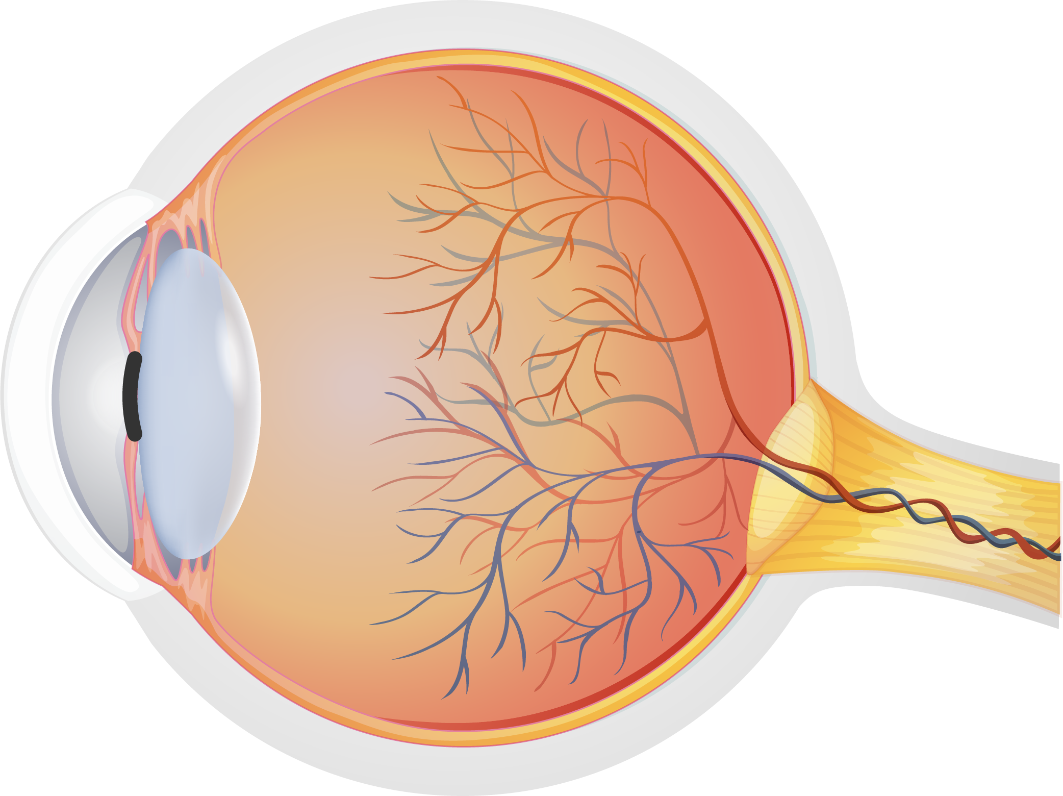
Healthy eye
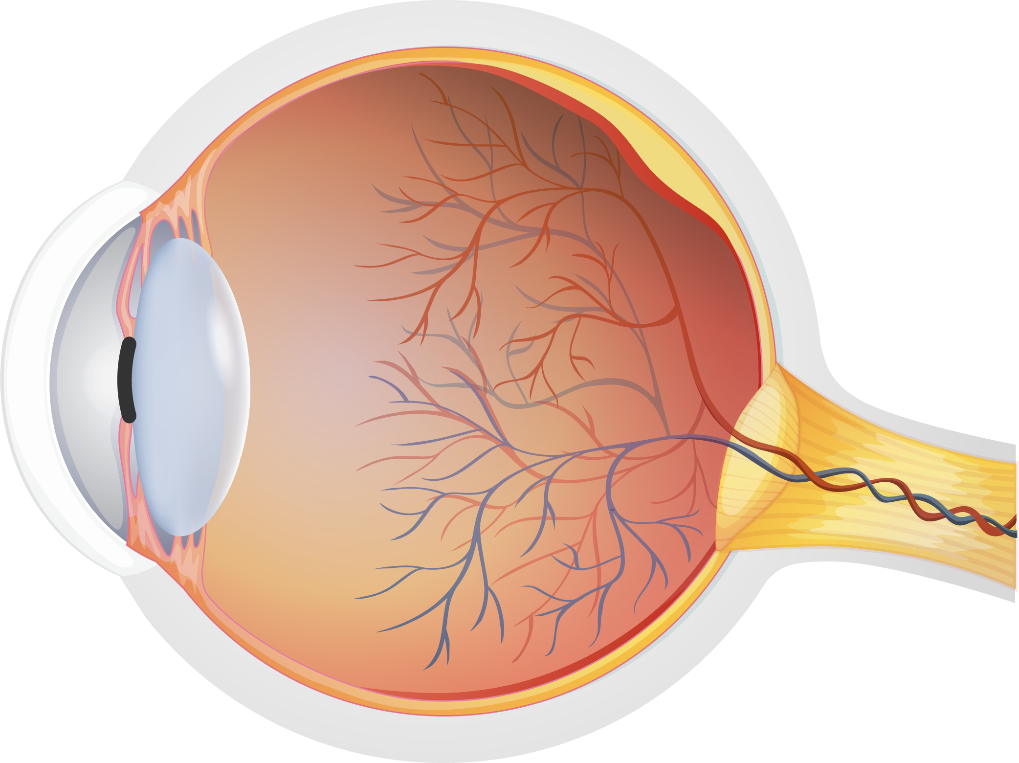
Retinal detachment
Symptoms
The following symptoms are early signs of retinal detachment. If any of these symptoms are present, it is necessary to call or visit an ophthalmologist immediately:
- Seeing floaters (myodesopias) in the form of spots, lines or spider webs in the visual field
- Seeing flashing lights
- The appearance of a peripheral shadow in the visual field or a dark curtain that gradually covers it
- Image distortion
Treatment
Retinal detachment is treated with surgery. There are several types of intervention, but these are the two most common types:
- Scleral surgery or scleral buckling: this consists of placing a silicone or soft plastic band on the sclera (white of the eye) with the purpose of compressing the retina towards the back of the eye to help reposition it. It is usually accompanied by cryocoagulation and laser photocoagulation techniques.
- Vitrectomy: this consists of removing the vitreous gel that sometimes presses on the retina, causing it to detach. The vitreous gel is usually replaced by a bubble of air, gas or oil, the purpose of which is to push the retina towards the back of the eye globe. This technique is usually accompanied by photocoagulation or cryocoagulation, which stimulates scarring to help reattach the retina.